
Updated June 2026 · 10-minute read

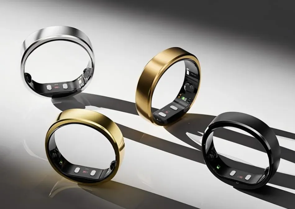
Atrial fibrillation detection was once a feature only available in hospitals, on clinical ECG machines operated by trained technicians. Today it runs passively inside a titanium ring on your finger, 24 hours a day, without any user action required. Oura Ring 4 and Samsung Galaxy Ring both carry FDA clearance for this capability.
What makes this remarkable is not just the hardware miniaturisation. It is the AI classification layer that converts optical light absorption data, not electrical signals from the heart, into a reliable screening signal for one of the most clinically important cardiac arrhythmias. This article explains exactly how that works.
Why AFib Is Hard to Detect Without an ECG
Atrial fibrillation is an electrical disorder of the heart. In normal sinus rhythm the sinoatrial node fires regular electrical impulses that trigger coordinated atrial contraction, then ventricular contraction, in a precise repeating pattern. In AFib the atria fire chaotically instead of coordinating. The ventricles still contract, driven by the atrioventricular node, but the timing of their contractions becomes irregular because they are responding to irregular atrial input.
The gold standard for detecting AFib is the ECG: electrodes placed on the skin measure the heart's electrical signals directly. The P wave, which represents atrial activation, disappears in AFib and is replaced by irregular fibrillatory activity. The RR intervals (time between successive ventricular beats) become irregular in a characteristic way. A trained cardiologist can read these patterns immediately.
A smart ring has no ECG electrodes. It has optical sensors that measure blood volume changes in the finger. The challenge for AI-based AFib detection is extracting enough information from this indirect signal to reliably identify the same rhythm abnormality that the ECG shows directly.
What the Optical Signal Looks Like During AFib
The PPG signal from the ring's optical sensor is fundamentally a measure of blood volume in the finger's capillaries with each heartbeat. Each heartbeat creates a pulse of increased blood volume that the sensor detects as a change in light absorption. The timing between these pulses (the inter-beat intervals) reflects the timing of the heart's ventricular contractions.
In normal sinus rhythm the inter-beat intervals are consistent and only vary slightly with breathing and other normal autonomic influences. This normal variation is what HRV measures. In AFib the inter-beat intervals are chaotically irregular: some beats come very close together, others are widely spaced, and there is no repeating pattern to the irregularity.
This difference in interval regularity is detectable in PPG data. The AI does not need to see P waves or exactly reconstruct the ECG. It needs to reliably detect that the inter-beat interval sequence has the characteristic irregularity pattern of AFib rather than the normal (or normally variable) pattern of sinus rhythm.
The AI Classification Pipeline
The AFib detection algorithm takes a sequence of inter-beat intervals extracted from the PPG signal and classifies the rhythm as either normal or potentially AFib. The specific pipeline used by Oura and Samsung is proprietary, but published research in this area describes the general approach that has achieved FDA clearance across multiple devices.
Step 1: IBI extraction and quality filtering
The first step is extracting clean inter-beat intervals from the raw PPG signal. Peak detection algorithms identify each heartbeat in the signal and record its timestamp. The interval between successive peaks is the IBI (inter-beat interval) sequence.
Not all peaks are reliable. Motion artefacts, poor skin contact, and other signal quality issues produce spurious peaks or missed peaks that would corrupt the IBI sequence and generate false classifications. The pipeline filters out IBI sequences where signal quality falls below a threshold. During movement, the AFib classification is suspended because the signal quality is insufficient. This is why AFib detection primarily runs during rest and sleep rather than during exercise.
Step 2: Rhythm regularity feature extraction
From a clean IBI sequence, a set of features is computed that characterises the regularity and pattern of the rhythm. Key features used in published algorithms:
RMSSD: The standard HRV metric, but used here differently. In sinus rhythm, RMSSD reflects normal autonomic variation. In AFib, RMSSD is elevated because of the chaotic irregularity, but the pattern of variation is different from normal HRV.
Coefficient of variation of IBI: The standard deviation of IBIs divided by the mean, capturing relative variability independent of heart rate.
Root Mean Square of Successive Differences normalised by mean IBI: Distinguishes AFib-type irregularity from high-HRV sinus rhythm.
Poincare plot features: A Poincare plot graphs each IBI against the next IBI. Sinus rhythm produces a compact elliptical cloud. AFib produces a diffuse, structureless scatter. SD1 and SD2 from the Poincare plot capture this shape difference.
Sample entropy: A measure of the complexity and unpredictability of the IBI sequence. AFib produces higher sample entropy than sinus rhythm because the irregularity has no repeating pattern.
Step 3: Classification
These features are fed into a binary classifier trained on labelled IBI sequences from patients with confirmed AFib and confirmed normal sinus rhythm. The classifier learns the feature combinations that reliably separate the two classes.
Logistic regression, support vector machines, random forests, and deep learning approaches (particularly 1D convolutional neural networks operating directly on IBI sequences) have all been used successfully for this classification. Published accuracy results for well-trained classifiers on clean PPG data typically show sensitivity (true positive rate for AFib) of 90 to 97 percent and specificity (true negative rate, avoiding false positives in sinus rhythm) of 90 to 96 percent.
The classifier output is a probability score rather than a binary decision. The threshold that converts probability to a notification is set conservatively to minimise false positives, which means some true AFib episodes may not reach the notification threshold. This is appropriate for a screening tool: a false positive causes unnecessary anxiety and a doctor visit, while a false negative delays detection. The threshold balancing is part of the FDA submission and review process.
What FDA Clearance Required
To receive FDA clearance for AFib notification, both Oura and Samsung had to submit clinical study data demonstrating that their algorithms achieved specified performance thresholds in populations both with and without confirmed AFib. The studies required:
Simultaneous recording of the wearable device and a clinical ECG reference in enrolled participants
A pre-specified performance claim (typically sensitivity above 90 percent and specificity above 85 percent)
Sufficient sample size to demonstrate performance with statistical significance
Testing in a population representative of the intended use population
The FDA's review assessed whether the study methodology was sound, whether the performance claims were met, and whether the labelling (how the feature is presented to users) was accurate about what the device does and does not do. The cleared indication specifies that the device notifies of irregular rhythm patterns that may be consistent with AFib, not that it diagnoses AFib. Diagnosis requires clinical confirmation.
Limitations the AI Cannot Overcome
Even a well-designed AI classifier operating on clean PPG data has inherent limitations for AFib detection.
Paroxysmal AFib. AFib that comes and goes (paroxysmal AFib) may not be present during the monitoring window when the ring is collecting sufficient quality data. A single normal overnight reading does not rule out paroxysmal AFib that occurs during daytime activity or at other times.
Other arrhythmias. Some other cardiac arrhythmias produce irregular IBI sequences that can look like AFib to an optical sensor-based classifier. Frequent premature atrial or ventricular contractions produce bigeminy or trigeminy patterns that may generate false positives in some algorithms. Clinical ECG is necessary to distinguish these.
Signal quality during movement. AFib detection requires clean PPG data, which means it primarily runs during rest and sleep. Active AFib episodes during exercise would not be reliably detected by current ring-based implementations.
Frequently Asked Questions
Can a smart ring replace a Holter monitor for AFib detection?
No. A Holter monitor records a continuous clinical-grade ECG for 24 to 48 hours and provides a complete record of all rhythm events during that window. A smart ring provides a screening signal that can prompt further investigation but cannot produce a clinical ECG record. For a patient with known AFib or suspected but unconfirmed AFib, a Holter monitor or extended cardiac event monitor provides far more diagnostic information than a consumer ring.
How does Apple Watch ECG compare to Oura Ring PPG for AFib detection?
Apple Watch uses a single-lead ECG that directly measures the heart's electrical signals, producing a rhythm strip that closely resembles a clinical ECG lead. Oura Ring uses optical PPG data and infers rhythm irregularity indirectly from inter-beat intervals. The ECG approach provides a more direct measurement of the underlying electrical abnormality. However, the PPG approach runs continuously and passively during sleep while the Apple Watch ECG requires a deliberate 30-second recording. Both have FDA clearance, and for passive screening during sleep the ring's continuous monitoring provides longer observation windows than the on-demand Apple Watch ECG.
What should I do if my smart ring flags a possible AFib episode?
Contact your healthcare provider and mention the notification. Do not ignore it, but also do not assume a diagnosis. The provider will likely order a clinical ECG or extended heart rhythm monitoring to confirm or rule out AFib. If you experience symptoms alongside the notification, such as palpitations, shortness of breath, or chest discomfort, seek prompt medical attention. If the notification appeared without symptoms and your provider confirms AFib, treatment options are available and AFib is highly manageable when detected early.
Why do smart rings only detect AFib and not other arrhythmias?
AFib is the most common arrhythmia and has a distinctive signature in optical PPG data (chaotic IBI irregularity) that is detectable without a clinical ECG. Other arrhythmias, such as ventricular tachycardia or heart block, have signatures that are more subtle or ambiguous in PPG data, require more leads of ECG data to characterise, or are rare enough that clinical training datasets are smaller. Regulatory clearance requires demonstrating performance in large enough clinical studies, which is most feasible for the most common condition. Research into PPG-based detection of other arrhythmias is ongoing.
Technical details draw on FDA 510(k) clearance documentation for Oura Ring 4 and Samsung Galaxy Ring, published research in IEEE Transactions on Biomedical Engineering, and peer-reviewed PPG-based arrhythmia detection literature as of June 2026.